
There are multiple reasons, but the main factor is the ability for your patients to see for themselves what you as their eye care professionals are telling them about the condition of their eye. This builds healthy patient behaviors as well as loyalty to your practice. Using a digital imaging system in your practice increases clarity not just in diagnosing your patients’ conditions, but also in communicating with others: your patients, your staff, and with doctors to whom you refer patients for tertiary care.
More and more ODs are turning to TelScreen’s EyeResTM slit lamp and imaging systems. Doctors Paul Karpecki, Ben Gaddie, and Mile Brujic, for example, are amongst the many ODs who have purchased and are presently using TelScreen products in their eye care practices.
What makes EyeRes digital imaging systems the best for your practice is a combination of the best high-performing technology, technical support, and client care that keeps ODs and Ophthalmologists returning to expand their practices with additional systems. To figure out what works best for your specific needs, give Wes a call at (502) 515-1806.
The benefits of utilizing a slit lamp digital imaging system in your eye care practice are for you and your patients. Your patients can tangibly see the eye conditions you are trying to explain to them and they become more compliant with recommendations. You create a stronger practice through patient referrals and an additional revenue source.
Short answer – it depends. While it’s not unusual to see anterior-segment imaging revenue of $600 per exam room per month, the best way to determine the right answer for you is to call Wes Harris at (502) 515-1806. He can walk you through the estimate of revenue in your practice based on your patient demographics, your most common 3rd-party payers, and your medical interests.
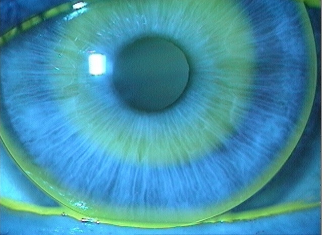
An eye-opening experience! The process is simple and with the ability to see for themselves what you as the professional view in a slit lamp, patients will have more confidence in you as their eye care provider. Videos and images help patients to understand more, and to understand more quickly. Any patient with a medical condition (keratitis, conjunctivitis, dry eye, etc) will benefit from a visual explanation, and will be more likely to adhere to your treatment plan. The EyeRes system is useful for contact lens fitting (send pictures to the lab for consultation) and for encouraging lens care & on-time replacement as well as other situations.
If you can see it through the slit lamp oculars, you can photograph it. For posterior photos at the slit lamp, a dilated pupil is extremely helpful, and a condensing lens is required. We recommend the Volk / TSi 1.0 Digital Imaging Lens.
When implemented correctly in a practice, the EyeRes system will not slow you down. With the expert advice of clinical advisors over the years, we have refined the user interface so that the use of a slit-lamp camera actually made the experience more efficient with the patient.
The photodocumentation procedures are defined by “what did you photograph” not by “which instrument did you use to photograph it.” Photos of the optic disk and macula are fundus photographs, regardless of how you acquired the image. You should bill for CPT 92250 Fundus Photography, after completing your I&R.
Seeing Is Believing
Let us help you show your patients what you see in them.